Is Your Pet’s Bump a Harmless Lump or a Tumor?
Discover how to differentiate between benign and malignant growths. Understand why recognizing the symptoms early and seeking veterinary advice can protect your pet from hidden dangers.

Reviewed by Dr. Becker


STORY AT-A-GLANCE
- When mast cells, which are found naturally in all the tissues of your pet’s body, replicate in abnormally high numbers, a mast cell tumor can be the result
- In dogs, mast cell tumors are most often found on the trunk, the limbs and between the toes; prognosis depends on the tumor location, the extent and grade of the tumor, and the type of treatment given
- Mast cell tumors are graded on a scale; a grade III or high-grade tumor is the most serious, with the worst prognosis for recovery
- If your pet is diagnosed with a mast cell tumor, it’s important to work with an integrative or holistic veterinarian who can offer a variety of complimentary therapies, including diet recommendations

Join the Bark & Whiskers™ Family
Sign up today for our FREE newsletter, packed with expert advice and insider tips to keep your beloved pet in tip-top shape.
View our Privacy Policy and Terms of Service.


Mast cell tumors (MCTs) are the most common type of skin cancer in pets and are much more prevalent in dogs than cats. These growths are the reason I always recommend careful monitoring of any skin growths on your canine companion, and alerting your veterinarian immediately if you find a new mass.
What to Look for When Good Mast Cells Go Bad
Mast cells exist in all the tissues of your pet’s body, but they’re found in the highest concentrations in the skin, respiratory tract and gastrointestinal (GI) tract. These cells are very rich in histamine, which is a vasodilator that promotes blood flow to tissues, and also heparin, an anti-coagulant that prevents blood from clotting too quickly. Both of these naturally occurring chemicals play a role in all allergic responses, non-allergenic skin disease, wound healing, and tissue remodeling. They can also increase stomach acid production.
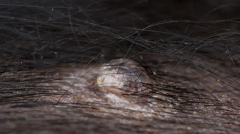
As with any cell in the body, when mast cells replicate in higher-than-normal numbers, a mast cell tumor can form. These tumors are actually quite common, accounting for about 20% of all skin tumors in dogs. If a pet has a mast cell tumor on the skin, there’ll be a bump or a lesion of some kind. Sometimes it’s a raised pink bump on the surface of the skin. It may be ulcerated, scabby, or oozing.
Mast cell tumors can be as small as a pencil eraser or as large as a softball, and importantly, they tend to wax and wane. They can start small and suddenly become large, red, and irritated or weepy. This is a sign of degranulation, meaning the tumor has become irritated and released the nasty substances within it. The hallmark of a mast cell tumor is a tumor that grows and shrinks periodically.
Sometimes the tumor will be a less-defined mass that feels like a lump under the skin, similar to a fatty lipoma. Because these tumors have the ability to mimic the appearance of many other types of skin problems, it’s very important to have your veterinarian check out any new lumps or bumps on your pet.
Dogs usually develop a single tumor (cats can develop multiple tumors), and although some may be benign, most mast cell tumors are cancerous (malignant).
Mast cell tumors can be irritating, so dogs will sometimes begin licking, scratching or picking at them. There can also be complications from the tumors, such as stomach problems from the overproduction of histamine and excessive bleeding from the release of heparin.
Diagnosing MCTs
Your veterinarian will perform a physical exam and focus on the mass. A fine need aspirate will likely be recommended; this involves using a very small needle to obtain a sample of cells from the tumor. Biopsy is another option. In either case, your vet may want to pre-medicate your dog with the antihistamine Benadryl, since mast cells contain histamine.
Giving Benadryl ahead of time may help prevent the tumor from degranulating during sampling. Sudden degranulation can cause a systemic reaction (anaphylaxis) that can be very serious — even life-threatening.
If the fine-needle aspirate reveals mast cells, it’s very important that the veterinary surgeon takes large margins around the tumor. This will reduce the likelihood of leaving tumor cells behind, which is unfortunately very common with mast cell resections. The tissue that is removed will be sent to a pathologist for staging or grading. This will let your veterinarian know how extensive the disease is and what type of treatment is needed.
Diagnosis is generally confirmed by a veterinary pathologist. There are two different methods for grading MCTs. The older method uses the Patnaik scale of I through III, with a rating of I designating the least malignant. The newer method is the Kiupel system, which simply distinguishes high grade vs. low grade.
Prognosis for Dogs With Mast Cell Tumors
Mast cell tumors in dogs are most often found on the trunk, the limbs and between the toes. The tumors are seen more often in certain breeds, including the Bulldog, Boston Terrier, Boxer, Pug, Labrador and Golden Retriever, Cocker Spaniel, Schnauzer, Staffordshire Terrier, Beagle, Rhodesian Ridgeback, Weimaraner, and the Shar Pei.
Prognosis depends on the tumor location, the extent of the tumor, the grade, and the type of treatment given. Mast cell tumors of the skin are very different in dogs than in cats. Surgery to remove the tumor is less invasive in cats, and the prognosis for a full recovery is much better in cats than in dogs.
Mast cell tumors with a generally poor prognosis are those on the muscle, in the mouth, in the internal organs, the bloodstream or bone marrow, and ulcerated tumors. Mast cell tumors that cause GI ulceration or are large, fast-growing or recurring also carry a much poorer prognosis.
Grade III tumors are the most serious and carry the worst prognosis for recovery. Grade I tumors generally have an excellent cure rate, as long as the entire thing is removed. And again, it’s important that the surgeon takes very wide margins.
If a dog goes 30 weeks post-surgery for a grade I tumor with no recurrence during that time, he’s considered cured.
Even with aggressive surgery, the recurrence rate for a grade II mast cell tumor is about 20%. The majority of dogs with grade III malignant mast cell tumors will experience spread of the tumor. Sadly, only about 10% of these dogs live longer than a year after surgery.
In addition to histopathology, which means looking at the cancer tissue microscopically, veterinarians also have the option to perform cell proliferation analysis through Michigan State University’s diagnostic lab, which is something I highly recommend. You can find more information at Michigan State University Canine Cutaneous Mast Cell Tumor Panel and MSU Mast Cell Tumor Flowchart.
This wonderful technology analyzes three markers to assess the risk of systemic disease. Pets with external tumors can have this additional test, which shows what’s going on inside the body. It can be very beneficial for the pet parent’s peace of mind as well as for prognosis planning.
Veterinarians using this technology to help formulate a treatment plan will have a much more accurate blueprint of what’s really going on with mast cells deep within the patient’s body. If your pet has been diagnosed with a mast cell tumor, I recommend that you ask for this additional test.
Additional Recommendations
I also recommend that you work with an integrative or holistic veterinarian to reduce the risk of recurrence, because these veterinarians use a variety of additional complimentary therapies that can be very beneficial, such as Ayurvedic medicine and Chinese herbs that naturally decrease the number of circulating mast cells in the body.
There are also nutraceuticals and additional supplements that can help naturally reduce mast cell degranulation and histamine release, which keeps the patient much more comfortable.
I also recommend that you eliminate any foods with carbohydrates if your pet has mast cell tumors, because carbohydrates create inflammation in the body. My best dietary suggestion is to institute a ketogenic diet for four months, as the anti-inflammatory effects can be profound. It’s what animals really need when their bodies are gearing up to fight off cancer or to heal from a surgical procedure. I also recommend supplementation with a rich source of omega-3 fatty acid, such as krill oil.
And I absolutely recommend if your pet has been diagnosed with mast cell tumors that you never vaccinate again. Additional vaccines can prompt a massive inflammatory response that can spur on additional mast cell tumors. From this point forward, if you have a pet diagnosed with mast cell tumors, I recommend that you titer.
Sources and References
Unlock Pro Tips for Pet Wellness
Get exclusive access to expert insights and holistic health tips for your furry friend – all for FREE because your pet’s well-being is our top priority.
View our Privacy Policy and Terms of Service.