
Cysts Versus Tumors — How to Recognize Lumps on Your Dog
See an unfamiliar growth on your pet's skin? Don't panic. Make an appointment with your integrative vet to determine what it is and if treatment is necessary.

Reviewed by Bark & Whiskers


Join the Bark & Whiskers™ Family
Sign up today for our FREE newsletter, packed with expert advice and insider tips to keep your beloved pet in tip-top shape.
View our Privacy Policy and Terms of Service.


STORY AT-A-GLANCE
- Lumps and bumps are common in dogs of all ages and breeds. Cysts are hollow sacs filled with fluid or semi-solid material, while tumors are solid masses of abnormally dividing cells
- Cysts are typically benign and non-contagious. There are five types — true cysts, false cysts, sebaceous cysts, follicular cysts, and dermoid cysts. They can vary in appearance and location on the body
- Tumors can be benign or malignant. Common types include mast cell tumors, melanomas, histiocytomas, and adenomas/adenocarcinomas. Some tumors may be mistaken for cysts and require veterinary attention
- Other growths that may appear on dogs include skin tags, papillomas (warts), and lipomas (fatty tumors). These are generally benign but may require removal if they’re causing discomfort
- It's important to have any new or changing lumps examined by a veterinarian. They will perform tests like biopsies or fine needle aspirations to determine if a growth is cancerous
It’s not uncommon to see a lump growing on your dog’s body — at any age and any breed, lumps and bumps are a common occurrence. However, it’s helpful to know the difference between a cyst and a tumor, and whether a sudden, unfamiliar growth is best left alone or needs to be removed.
What Is a Cyst?
Cysts are a type of hollow sac found on a dog’s skin, which is then filled with either a fluid, semi-solid or solidified material. These contents are usually natural body secretions from the dog, such as sebum (oil substance secreted by the sebaceous glands) or breakdown products like dead skin cells and keratin.1
Some cysts are small, while others are large. They appear in various areas of the body, such as under the skin of the head, neck, trunk and eyelids. Sometimes, they grow in between the toes.2 According to Jerry Klein, chief veterinary officer for the American Kennel Club, although cysts can develop within the tissue or in any part of the body, most of the cysts we think of are found on or just under the skin.3
Different Types of Cysts in Dogs
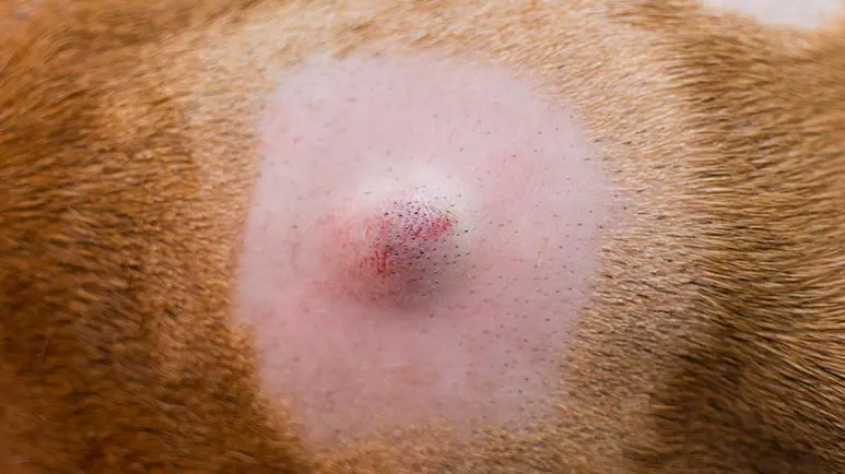
Cysts can look and feel different, depending on the type. Usually, these are slow-growing, smooth, raised bumps that ulcerate, change color and ooze discharge. Cysts often appear white, blue or a dark color.
Don’t worry if one of your dogs has a cyst and the other doesn’t. These lumps are not contagious between dogs. These growths are also benign, meaning they aren’t cancerous, and do not spread to other parts of the body. There are several types of cysts, which are divided into five kinds:4,5
- True cysts — In dogs, true cysts usually form around the eyes or in their ears. These are usually associated with the sweat glands, and the lining often secretes fluid — the more fluid secreted by the lining, the bigger the bump becomes.
True cysts usually appear translucent, dark or bluish. They leak a yellowish material. According to Klein, true cysts are usually surgically removed by vets to prevent a recurrence. - False cysts — As opposed to true cysts, these do not have a lining that secrete fluid. Rather, they are formed because of a trauma or injury, and contain dead tissue that liquifies, leading to a dark, fluid-filled mass.
- Sebaceous cysts — These cysts occur near hair follicles, and form when an oil gland becomes blocked. Hence, the growths are usually filled with sebum and have a whitish or bluish appearance. Sebaceous cysts can break open or bleed, which makes them prone to infections.
- Follicular cysts (epidermoid cysts) — Klein says that these cell cysts are “associated with the base of the hair follicle, which often becomes irritated or inflamed.” These cysts are usually hard and are filled with different types of materials. When expressed and pushed out, the discharge is either black, white or have a foul-smelling, cheesy appearance.
In dogs, follicular cysts appear around the legs or mouth. Just like sebaceous cysts, they are prone to infection. - Dermoid cysts — These cysts form when the epidermis separates from the underlying tissue. They rarely occur in dogs, but when they do, they usually manifest in two dog breeds — the Rhodesian Ridgeback and Kerry Blue Terrier. Dermoid cysts appear on the dog’s neck and are congenital, although they may not appear until the dog is 9 to 12 months old.
What Is a Tumor?
Compared to cysts, tumors require more urgent attention. These solid masses are made up of cells that divide abnormally. Instead of following the natural cell cycle that ends in cellular death, the abnormal cells multiply out of control. This occurs when the cells’ DNA is damaged due to various factors, leading to the abnormal mutation.
Tumors can be either benign or malignant. Malignant means they are cancerous, and can spread to other areas of the body. Depending on the type and location, tumors can lead to different symptoms.6,7
Common Types of Tumors in Dogs
Below are some examples of tumors that are often mistaken for cysts:8,9
- Mast cell tumors (MCTs) — These are the most common type of skin cancer in dogs. They grow below or on the skin, and vary in size and appearance — some are soft, while others are firm. MCTs can be hairless, ulcerated, raised or discolored. According to Dr. Julie Buzby:
“[M]ast cells, which are part of the immune system, are filled with histamine granules. Therefore, squeezing or otherwise aggravating a mast cell tumor may cause the mast cells within to degranulate, releasing large amounts of histamine. This can cause anaphylaxis (i.e. a serious allergic reaction).”
- Melanomas — These growths result from melanocytes, which are the pigment-carrying skin cells. In dogs, melanomas usually occur in their oral cavity, although these growths can manifest in their eye, nail bed and trunk. They are usually pink or non-pigmented, and are flat as opposed to raised.
Melanomas on the skin are usually benign, but on the nail bed and oral cavity, these growths are malignant. Specifically, oral melanomas can be darkly pigmented or pink, and a common symptom is having bad breath. - Histiocytomas — Young dogs are prone to getting these pink, round, raised and sometimes ulcerated lumps. Histiocytomas are sometimes mistaken for MCTs, however, they are benign and do not need to be removed or treated; rather, they are eliminated by the dog’s immune system.
- Adenomas or adenocarcinomas — These small, irregularly shaped and sometimes ulcerated growths come from the sebaceous glands. When benign, these masses are called adenomas, but when malignant, they are called adenocarcinomas. Adenocarcinomas can start out small but grow bigger as they progress.
Other Growths That May Appear on Your Dog’s Body
Remember that not all growths that you see on your dog’s body may be cysts or tumors. There are other types that you need to identify as well. Here are some examples:10
- Skin tags — These are similar to the tags that appear on people’s body. They can get big and hang off the skin by a narrow stem. Skin tags do not need to be removed, unless they are causing discomfort to your dog.
- Papillomas — These are warts that appear on your dog’s lips, face and inside the mouth. Like cysts, these are benign; however, unlike cysts, they are very contagious. Papillomas usually resolve on their own, but if they are causing problems for your pet, they can be removed.
- Lipomas — These are fat-filled, solid benign tumors that are found under the fatty layer of your dog’s skin. Usually, lipomas are a cosmetic issue and are not dangerous. However, if located in an area where it hampers movement, such as on the legs, a lipoma may be removed by your vet.
Bring Your Dog to the Vet to Have the Lump Checked
Most pet parents panic at the first sight of an unknown growth on their dog’s body. However, the best thing to do is to consult with an integrative vet to help diagnose the lump. It’s advisable to observe the growth — check if it changes color, grows bigger or becomes inflamed.
If the mass is oozing, inflamed or painful to the touch, don’t delay the checkup, as it could require emergency treatment. After a physical examination, your vet may do certain procedures like a biopsy or a fine needle aspiration to confirm if the growth is cancerous or not. The key is to work with your vet to devise a treatment plan to help excise the lump (if needed) and manage the condition.