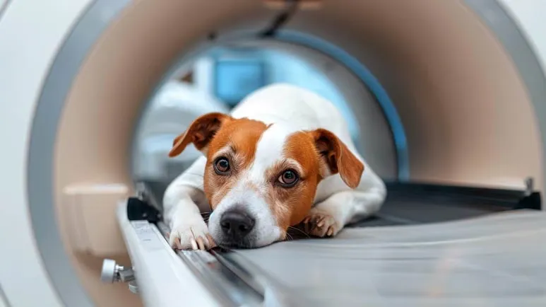
What Health Secrets Could Be Lurking in Your Dog's Brain Scans?
An MRI scan can be helpful to identify neurological disorders, but it isn't foolproof. These studies show how this diagnostic tool can help identify diseases in pets.

STORY AT-A-GLANCE
- Magnetic resonance imaging (MRI) technology, commonly used in human medicine for internal organ imaging, has valuable applications in veterinary medicine, particularly for diagnosing neurological disorders in pets through brain scans
- An earlier study of dogs with Granulomatous Meningoencephalomyelitis (GME) showed MRIs have limitations, as they failed to detect meningeal inflammation in dogs with this illness
- However, recent research demonstrates MRIs can identify changes in brain chemicals (biomarkers) like NfL, Tau Protein, NSE and Amyloid-β, helping diagnose conditions such as brain tumors and meningoencephalitis
- A study of 73 dogs with MUE revealed that those with regular MRI scans had significantly better survival rates than those showing visible damage, with only 5% mortality versus 33%
- While MRI technology has limitations, it remains a valuable diagnostic tool in veterinary medicine, helping determine disease severity and guide treatment plans for pets with neurological conditions
Diagnostic tools like magnetic resonance imaging (MRI) are widely used in human medicine, providing detailed images of the internal organs to determine medical conditions, including diseases and tumor growth. But did you know these tools can also be helpful in veterinary medicine?
In particular, using MRIs to scan the brain can help diagnose neurological disorders in your animal companions. This article will highlight three animal studies on veterinary uses of MRIs and how they can help identify brain issues in your pets.
MRIs Are Useful but Not Foolproof, an Earlier Study Finds
The first study to be discussed dates back to 2006.1 Published in The Veterinary Record journal, it looked at the MRI scans of the brain and spinal cords of 11 dogs diagnosed with Granulomatous Meningoencephalomyelitis (GME). This severe inflammatory disease occurs when the immune system attacks the dog’s nervous system, leading to swelling and damage.
According to their observations, while MRIs could identify areas of the brain and spinal cord that were damaged or inflamed, there are instances where the scans miss something.
For example, the MRI did not show any noticeable changes on the meninges, which are the layers that cover the brain and spinal cord. Still, when tissue samples were analyzed, they found that most dogs had inflammation in their meninges. Injecting a contrast dye (used to improve the quality of the scans) helped the GME-associated lesions stand out, but not always.
Overall, while MRIs can identify lesions, the appearance of these lesions in GME appears similar to other diseases. Hence, an MRI alone cannot definitively diagnose GME and may need other tests and clinical information to confirm the diagnosis.
MRIs Can Help Identify Changes in Brain Chemicals, Which Indicate Disorders
More recent studies, however, highlight the usefulness of MRIs in identifying brain chemicals, which can be a more reliable method in diagnosing brain and spinal cord diseases in dogs. These chemicals, found in the cerebrospinal fluid (CSF) in the brain and spinal cord, act as "biomarkers" to identify any damage.
Published in the journal Science Reports,2 the study involved dogs suffering from brain tumors, meningoencephalitis of unknown origin (MUO), and selected noninfectious myelopathies. The researchers acquired CSF samples and looked at four biomarkers, namely neurofilament light chain (NfL), tau protein, neuron-specific enolase (NSE), and amyloid-β protein.
They found that dogs with a specific health disorder had certain biomarkers that were either elevated or decreased. For example, in patients with MUO, NfL, NSE, and tau protein levels were high, indicating brain damage, while amyloid-β was lower than usual. In spinal cord disorders, no changes were seen in the biomarkers.
While the researchers note that looking at these biomarkers may not be sufficient for screening, they have "potential in improving the diagnostic precision."
"The combination of NfL, tau, and NSE may represent useful biomarkers for MUO as they reflect the same pathology and are not influenced by age …
Future studies are warranted to assess their prognostic and theragnostic utility in the treatment of canine MUO," the study authors concluded.3
Diagnostic Tools Can Help Identify Survival Rate in Diseased Dogs
We will discuss the final study, which the Journal of Veterinary Internal Medicine published,4 looked at the survivability rate of dogs with Meningoencephalomyelitis of Unknown Etiology (MUE) based on whether or not their MRI scans show brain or spinal cord damage. The researchers aimed to answer these questions:
- Do dogs with regular scans survive longer than dogs with abnormal scans?
- Can MRI findings help predict a dog’s chance of survival with MUE?
To conduct their research, the authors looked at 73 dogs with MUE, dividing them into two groups — one group had regular MRIs (without visible signs of damage). In contrast, another had abnormal scans (visible signs of damage).
The researchers found that dogs with regular MRIs lived longer than those with abnormal MRIs. The dogs with abnormal MRIs also had a higher mortality risk, with 33% of them succumbing to the disease (versus only 5% in the normal MRI group).
"Based on our findings, the presence or absence of MRI lesions in MUE dogs is prognostically relevant and suggests a potential role for MRI as a biomarker of disease severity," the researchers reported.5
The takeaway is apparent — dogs affected by this disease that show regular MRI scans have a much better chance of surviving than those with visible brain or spinal cord damage. This highlights the usefulness of MRIs in predicting the severity of neurological conditions and guiding treatment plans.
MRIs Can Help Diagnose Pets
These three studies underscore the advantages of using advanced imaging techniques and biomarker analysis to diagnose complex neurological diseases in dogs. They can also help determine the prognosis and treatment plan for your pet.
Hence, it’s vital to understand the strengths and limitations of this imaging tool to improve clinical outcomes and guide progressive research in veterinary medicine. If your pet undergoes an MRI, discuss the results with your integrative veterinarian so you can work together to help your pet recover.


